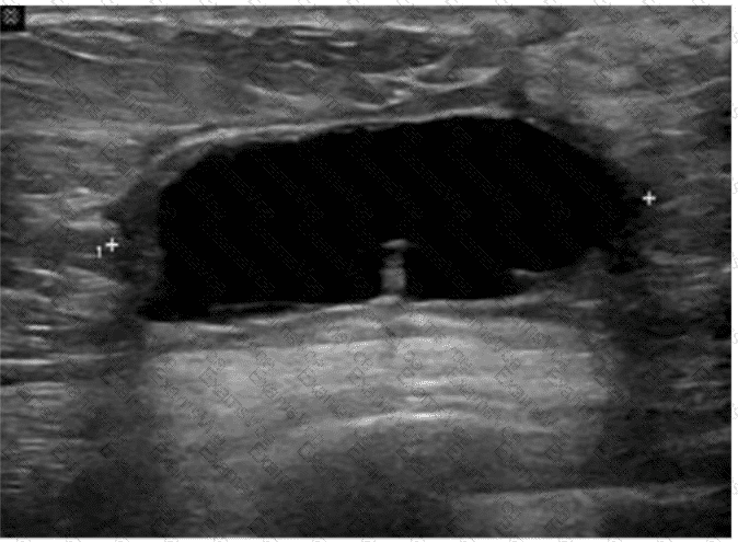
The ultrasound image reveals a well-defined, anechoic (black), thin-walled fluid collection located in the subcutaneous or parenchymal plane of the breast. This is most consistent with a seroma, particularly in the context of recent breast surgery.
A seroma is a common postsurgical finding, representing a sterile collection of serous fluid that accumulates in the surgical bed. It typically appears:
Anechoic (or hypoechoic if older)
Well circumscribed
Without internal septations or debris
Lacking hyperemia or surrounding inflammatory changes
This contrasts with:
A. Carcinoma — typically presents as an irregular, hypoechoic mass with angular margins, internal vascularity, and shadowing.
B. Blood clot (hematoma) — often appears heterogeneous, with internal echoes and variable echotexture depending on the age of the clot.
C. Abscess — appears as a complex fluid collection with thick walls, internal debris, septations, and surrounding hyperemia (often with clinical signs of infection).
D. Seroma — Correct. The described anechoic, clean-walled fluid collection is classic for a postoperative seroma.
[References:, Mendelson EB, Böhm-Vélez M, Berg WA.ACR BI-RADS® Atlas: Ultrasound. American College of Radiology; 2013., Stavros AT. Breast Ultrasound. Lippincott Williams & Wilkins; 2004., Rumack CM, Wilson SR, Charboneau JW, Levine D. Diagnostic Ultrasound, 5th ed. Elsevier; 2017., , ]