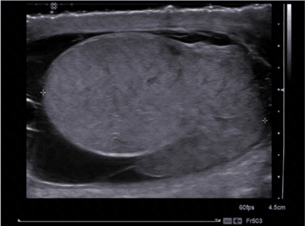
The grayscale ultrasound image demonstrates a uniformly enlarged, hypoechoic (dark), and heterogeneous testis without signs of surrounding scrotal wall thickening or a discrete fluid collection. This pattern is highly suggestive of testicular torsion in the setting of acute scrotal pain.
Sonographic features of testicular torsion on grayscale imaging:
Enlarged testis
Diffusely hypoechoic parenchyma
Loss of normal homogeneity
Absence of internal vascular flow on Doppler imaging (not shown here but critical in confirming diagnosis)
Testicular torsion occurs due to twisting of the spermatic cord, leading to vascular compromise and eventual infarction if not promptly corrected. It is a surgical emergency and typically presents in adolescent males with sudden-onset, severe unilateral testicular pain.
Comparison of answer choices:
A. Scrotal abscess appears as a complex fluid collection with irregular margins and posterior enhancement.
B. Testicular rupture would show discontinuity of the tunica albuginea, heterogeneous texture, and often a hematocele.
C. Testicular torsion — Correct. The enlarged, hypoechoic, heterogeneous testis is characteristic, particularly in the acute phase.
D. Epididymitis typically shows an enlarged, hypervascular epididymis and may extend to the testis (epididymo-orchitis), but vascularity is usually increased rather than absent.
[References:, Dogra VS, Gottlieb RH, Oka M, Rubens DJ. Sonography of the scrotum. Radiology. 2003;227(1):18–36., Rumack CM, Wilson SR, Charboneau JW, Levine D. Diagnostic Ultrasound, 5th ed. Elsevier; 2017., AIUM Practice Parameter for the Performance of a Scrotal Ultrasound Examination (2021)., , ]