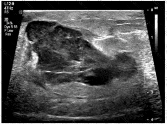
The ultrasound image demonstrates a complex, heterogeneous hypoechoic collection within the abdominal wall, with mixed echogenicity and ill-defined margins. The lesion appears to contain internal debris but lacks definitive signs of vascularity or air (which would be seen in an abscess). There is no peristalsis, herniated bowel, or fat to suggest hernia.
Given the history of atrial fibrillation — a condition commonly treated with anticoagulation therapy (e.g., warfarin, apixaban) — this clinical background raises high suspicion for a rectus sheath or abdominal wall hematoma.
Key ultrasound features of hematomas:
Early (acute): hyperechoic or heterogeneous
Chronic/resolving: complex or cystic with fluid-debris levels
No internal vascularity on Doppler
May be confined to muscle or fascial planes
This is consistent with a hematoma, particularly in patients on anticoagulation therapy.
Comparison of answer choices:
A. Hernia — typically shows bowel or fat with movement/peristalsis passing through a fascial defect.
B. Lipoma — usually homogeneous and echogenic, not complex or fluid-filled.
C. Abscess — often presents as a complex fluid collection with peripheral hyperemia and possibly air, plus systemic signs of infection.
D. Hematoma — Correct. The image and clinical history (anticoagulation due to atrial fibrillation) strongly support this diagnosis.
[References:, Berman L, et al. Sonographic appearance and evolution of rectus sheath hematomas. AJR Am J Roentgenol. 1996., Rumack CM, Wilson SR, Charboneau JW, Levine D. Diagnostic Ultrasound, 5th ed. Elsevier; 2017., AIUM Practice Parameter for the Performance of Diagnostic Ultrasound Examinations of the Abdomen and Retroperitoneum (2020)., ]