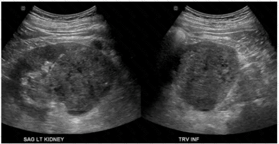
The ultrasound images (sagittal and transverse views of the left kidney) demonstrate a large, well-defined, heterogeneous mass within the renal parenchyma. This is highly characteristic of renal cell carcinoma (RCC), the most common primary renal malignancy in adults.
Renal cell carcinoma accounts for approximately 85% of all malignant renal tumors in adults. RCC often appears as:
A solid, heterogeneous, hypoechoic to isoechoic mass within the kidney
May contain areas of necrosis or hemorrhage (seen as mixed echogenicity)
Distortion of the normal renal contour
May have internal vascularity on Doppler imaging
Clear cell carcinoma (choice B) is the most common histological subtype of RCC but is not a separate diagnosis from RCC in imaging terms. Therefore, the most accurate answer is choice C: Renal cell carcinoma.
Differentiation from other options:
A. Nephroblastoma (Wilms tumor): A pediatric renal tumor, typically seen in children under 5 years of age—not applicable in adults.
B. Clear cell carcinoma: Histological subtype of RCC, not a distinct radiologic diagnosis.
D. Transitional cell carcinoma: Arises from the renal pelvis or ureter, typically appears as a central or collecting system mass rather than a cortical/parenchymal one.
[References:, Rumack CM, Wilson SR, Charboneau JW, Levine D. Diagnostic Ultrasound. 5th Edition. Elsevier, 2018. Chapter: Kidneys, pp. 215–222., Radiopaedia.org. Renal cell carcinoma: https://radiopaedia.org/articles/renal-cell-carcinoma, American College of Radiology (ACR) Appropriateness Criteria® – Hematuria, 2022., , , ]